
Live recordings of cell communication
A new advanced method for nano-scale imaging of
vesicle-fusion - vesicles are biological nano-sized
containers - could add to our understanding of diseases of
the nervous system and viral infections. In the long term,
this could be useful in developing a cure for neurological
diseases and mental disorders (e.g. schizophrenia,
depression, Parkinson's disease, Alzheimer's disease).
Researchers from the Department of Neuroscience and
Pharmacology and the
Nano-Science Center at the University of Copenhagen are
behind the new data, which have recently been published in
the prestigious
scientific journal PNAS.
Neurons communicate with each other with the help of nano-sized vesicles. Disruption of this communication process is responsible for many diseases and mental disorders like e.g. depression. Nerve signals travel from one neuron to another through vesicles - a nano-sized container loaded with neurotransmitter molecules. A vesicle fuses with the membrane surrounding a neuron, releases neurotransmitters into the surroundings that are detected by the next neuron in line. However, we still lack a more detailed understanding of how the fusion of vesicles occurs on the nano-scale.
Associate Professor Dimitrios Stamou, Department of Neuroscience and Pharmacology and Nano-Science Center explains:
- Contact between vesicles and membranes are an essential step in many important biological processes. We can now quantify contact areas formed between vesicles and determine the vesicle size and shape with nano-scale resolution. This helps us characterise the properties of the molecules involved in vesicle-fusion. The new method opens great new prospects for the research of neurological and infectious diseases.
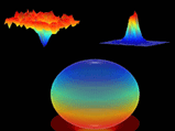
Researchers determine shape and size of the contact area between vesicle and membrane by measuring colour intensity from flourescent molecules. Right: Vesicle marked by acceptor flourescent molecules that light up when close to donor molecules (left). Middle: A plot of the same, calculated FRET.
Images on the nano-scale
The researchers are using a method called FRET (Fluorescence Resonance Energy Transfer). The method is well
known, but what is new is the way the researchers are using
it. They produce vesicles in the laboratory, which contain
fluorescent donor molecules, and membranes fixed to a
surface. The fixed membranes contain acceptor fluorescent
molecules. Only when the two different fluorescent molecules
are near to each other will light be emitted, which
researchers can measure as a sign of vesicle fusion. By
measuring the emitted light the researchers found new ways
to determine the vesicle shape with nano-scale resolution in
real-time.
- We have lacked a method for measuring the fusion of vesicle and membrane on a nano-scale at the moment the process occurs. Until now it has only been possible to get a still image of the process with high resolution, or live images with low resolution. With the new method we can quantify the changes in vesicle shape live i.e. during fusion, and with nanoscale resolution, explains Dimitrios Stamou.
More information:
Associate Professor Dimitrios Stamou
Email:
stamou@nano.ku.dk, +45 35 32 04 79 or +45 41 16 04 68
Communication Officer Gitte Frandsen
Email: gf@nano.ku.dk, +45 28 75 04 58
Link to the article in PNAS.